
Figure 3.
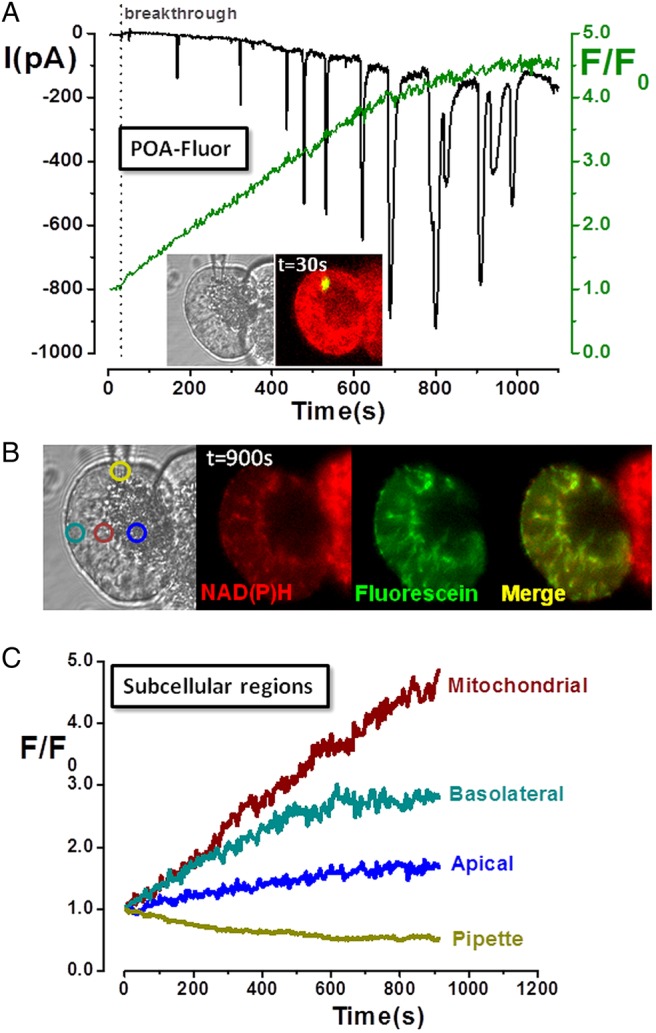
Mitochondrial localisation and activation of fatty acid ethyl ester probe (palmitoleic acid (POA)-Fluor) in pancreatic acinar cells. (A) Light-transmitted and fluorescent images of acinar cell doublet (inset) at time of membrane rupture (breakthrough) show localised fluorescence at patch-pipette tip as POA-Fluor activation by hydrolases released fluorescein. Time-dependent fluorescence rise (green) was associated with inward Ca2+-activated Cl− currents (IClCa), whereas no fluorescence was detected in the adjacent cell. (B) Light-transmitted and fluorescence images, and (C) subcellular regions of interest show predominantly mitochondrial distribution of fluorescence (fluorescein:NAD(P)H co-localisation), consistent with mitochondrial probe activation. [Ca2+]C levels increased over time (sustained inward IClCa with superimposed transients), accompanied by a concomitant decrease of NAD(P)H (not seen in non-patched, adjacent cell), consistent with fatty acid-induced mitochondrial inhibition.