
Figure 2.
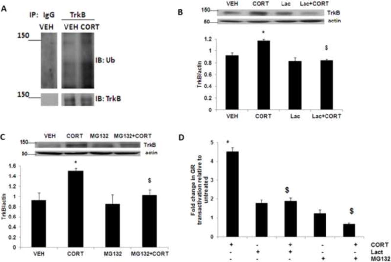
Acute corticosterone treatment induces TrkB ubiquitination. (A) Corticosterone treatment induces TrkB ubiquitination in neurons. Primary cortical neurons (DIV 4) were treated with vehicle (VEH) or corticosterone (CORT) for 3 h and lysates were immunoprecipitated (IP) with an anti-TrkB antibody, followed by immunoblotting (IB) with an anti-ubiquitin antibody (Ub) or anti-TrkB antibody. IgG, IgG control. Results are representative of three independent experiments. Primary cortical neurons (DIV 4) were treated with CORT or CORT plus the selective proteasome inhibitor (B) lactacystin (Lac) or (C) MG132, and lysates were subjected to immunoblot analysis. The upper panel shows representative autoradiogram of TrkB and actin, and the lower panel represents fold change in normalized TrkB protein levels. Results are mean ± SE for at least three independent preparations. *p<0.05 vs VEH; $p<0.05 vs CORT. One-way ANOVA followed by post-hoc Dunnett test. (D) Primary cortical neurons at DIV-4 were transiently transfected with the plasmids pHHLUC and pCMVβgal. Twenty-four hours after transfection, cells were treated with CORT or CORT plus the selective proteasome inhibitor lactacystin (Lac) or MG132, and luciferase activity was measured. Results are mean ± SE for at least three independent preparations expressed as fold change in GR transactivation relative to untreated cells. *p<0.05 vs VEH; $p<0.05 vs CORT. One-way ANOVA followed by post-hoc Dunnett test.