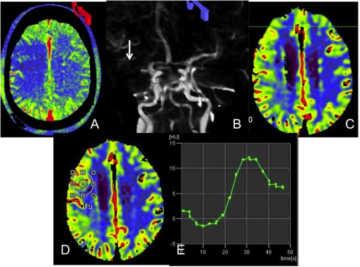
Figure 3.
A 72-year-old with an occluded right middle cerebral artery (MCA) presenting with an acute right hemisphere syndrome. Axial reconstructions from the flat detector-parenchymal blood volume (FD-PBV) examination demonstrate a large right frontal lobe defect (A). Reconstructed source data from the FD-PBV examination confirm the right MCA M1 segment occlusion (B, arrow). Conventional CT derived cerebral blood volume (CBV) examination demonstrates no deficit within the right MCA territory (C). A region of interest placed on the conventional CT CBV map corresponding to this right frontal region (D) generates dynamic parenchymal enhancement curves which peak approximately 30 s after the initiation of imaging (E). FD-PBV imaging is initiated, on average, 20 s after the start of the contrast infusion, well before saturation of PBV. This example demonstrates the capacity for PBV maps to overestimate CBV deficits in the setting of a proximal large vessel occlusion, particularly in an older patient with compromised cardiac output and delayed flow.

