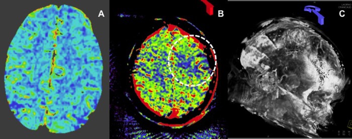
Figure 4.

A 76-year-old patient presenting with a symptomatic right carotid stenosis. Conventional CT derived cerebral blood volume maps demonstrate no deficits within the left hemisphere (A). MR imaging (not shown) confirmed that there was no stroke within the distribution of the left hemisphere. Flat detector-parenchymal blood volume (FD-PBV) map shows a wedge shaped PBV defect within the left anterior frontal lobe (B). Evaluation of the reconstructed source images demonstrates substantial patient motion with a significant misregistration of the native and subtracted post-contrast imaging, resulting in visualization of the soft tissue and bone structures on the three-dimensional reconstruction (in properly registered studies, these structures are subtracted out) (C).
