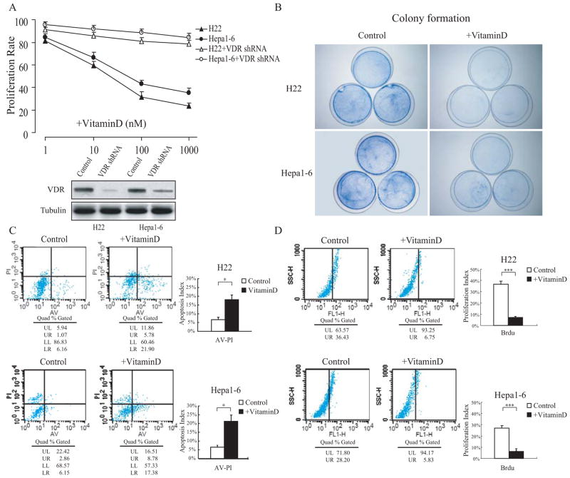
Fig. (1). 1,25(OH)2D3 inhibits cell proliferation and induces apoptosis in HCC cells.
The mouse-derived HCC cell lines H22 (Babl/c background) and Hepa1–6 (C57BL background) were different concentrations of 1,25(OH)2D3 the day after plating, and the effects were observed 48 hours later. (B) Colony formation was performed to assess cell proliferation in vitro. (C) H22 and Hepa1–6 cells were incorporated with 1μM BrdU to the culture medium 28 hours after 1,25(OH)2D3 or vehicle treatment, and cell proliferation analysis was performed by FACS. (D) Apoptosis analysis by Annexin V and PI double staining was performed by FACS. The results are representative of at least 3 independent experiments. *p < 0.05, ***p < 0.001.

