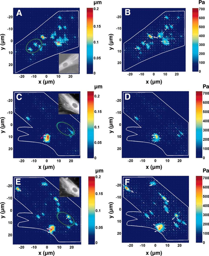
Fig. 6. Prestress dictates force propagation in the living cell.
A) and B): A normal smooth muscle cell displacement (A) and stress (B) maps, exhibiting long-distance force propagation behavior (inset in (A), YFP (yellow fluorescent protein)-actin image of the cell). C) and D): Long-distance force propagation disappears (loss of displacements and stress concentration spots away from the loading site, the magnetic bead) after inhibition of prestress by overexpressing caldesmon. Displacement and stress fields of a cell whose prestress was inhibited by being infected with a low level of green fluorescent protein (GFP)-caldesmon. E) and F): Long-distance force propagation resumes after caldesmon is inhibited. Displacement and stress maps of the same cell in (C) and (D) after treatment with calcium ionophore A-23187 (5 µg/ml for 10 min), an inhibitor of caldesmon. The pink arrow, bead direction and displacement magnitude. Note that when prestress is downregulated (inset in C) or is resumed (inset in E), there are no apparent changes in patterns of stress fibers compared with those in a normal cell (inset in A). Insets in C, and E are fluorescent images of the corresponding cell. Green ellipses represent the position of the nuclei. (Reprinted with permission from Hu et al 2003).

