
Figure 4.
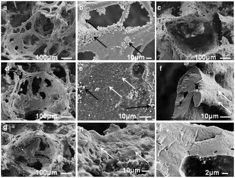
Scanning electron microscope (SEM) images of aqueous-derived scaffolds (6% w/v matrix, 500–600 μm NaCl particles); (a) with no reinforced silk particles, (b) magnified image from “a,” (c) reinforced with 24% PCR-AM particles, (d) reinforced with 24% PCR-AM-AJM particles, (e) magnified image of pore wall of “d,” (f) magnified image of fracture surface of “d,” (g) reinforced with 24% BM-AM particles, (h) magnified image of pore wall of “g,” (i) magnified image of fracture surface of “g.” Black arrows show the self assembled silk particles formed due to salt-matrix silk interactions. White arrows show the location of the embedded reinforced silk particles.