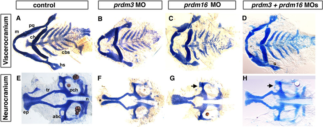
Fig. 2.
Reduction in prdm3 and prdm16 results in craniofacial defects. Five-dpf uninjected (A, E), prdm3 (B, F), prdm16 (C, G), and double morphant prdm3 and prdm16 (D, H) larvae stained with alcian blue to detect cartilage following dissection of the viscerocranium and neurocranium. As compared to control (A, E), the flat-mounted 5-dpf prdm3 i2e3 (B, F) and prdm16 e3i3 (C, G) morphant larvae have smaller palatoquadrate, including the pterygoid process of the palatoquadrate, and hyosymplectic of the viscerocranium, shortening of the Meckel’s cartilage (m), widening of the angle between ceratohyals (ch) in the viscerocranium, smaller ethmoid plate (ep), shortened trabeculae of the neurocranium, thinning and smaller of the neurocranium. In prdm3 morphant larvae (20–30%), there is a gap in the anterior edge of the ethmoid plate forming a “cleft” as shown in F. D, H: The combination of sub-optimal doses of prdm3 with prdm16 Morpholino. As compared to control (A, E), combination of 3 ng prdm3 i2e3 with 5 ng prdm16 e3i3 Morpholino (D,H) resulted in a slightly more severe phenotype in the neurocranium (much smaller and thinner neurocranium, smaller ethmoid plate, shortened trabeculae, missing anterior basicapsilar commissure, arrow). However, there is only a small gap in the ethmoid plate. Anterior is to the left. abc, anterior basicapsular commissure; cbs, ceratobranchials; ch, ceratohyal; ep, ethmoid plate; hs, hyosymplectic; m, Meckel’s cartilage; n, notochord; pch, parachordal; pq, palatoquadrate; tr, trabeculae; * indicates cleft in the ethmoid plate.

