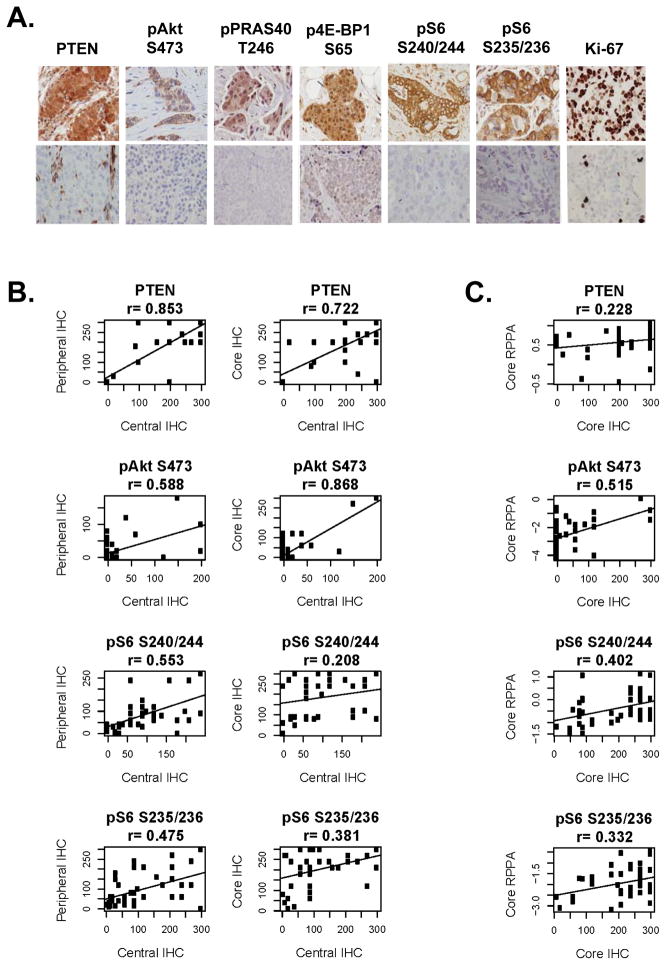
Figure 5.
IHC for PI3K markers. A. IHC for PTEN, pAkt S473, pPRAS40 T246, p4E-BP1 S65, pS6 S240/244, and Ki-67. Examples of positive (top panel) and negative (lower panel) immunostaining for each marker are included. B. Correlation of immunostaining for PTEN, pAkt S473, pS6 S240/244, and pS6 S235/236 between central and peripheral specimens (left), and central specimens and core needle biopsy (right). Immunostaining was quantitated by H-score. C. Correlation of central RPPA results with core needle biopsy specimen immunostaining for PTEN, pAkt S473, pS6 S240/244, and pS6 S235/236.

