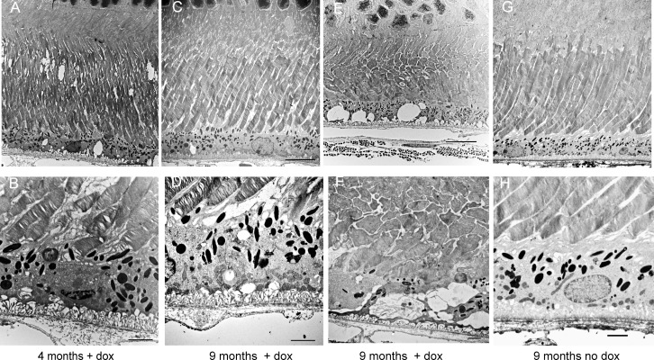
Figure 9.
Ultrastructural evidence of the RPE and photoreceptor atrophy. Electron micrographs were prepared at low (top) and high (bottom) magnification (scale bars: 2 μm) from eyes of 4-month-old and 9-month-old Sod2flox/floxVMD2cre mice with or without doxycycline induction. (A, B) Even at 4 months, the photoreceptor outer segments appeared disordered in doxycycline-induced mice, and basal infoldings of the RPE were disordered. (C, D) By 9 months, disc membranes were more widely spaced, and fragmented mitochondria had accumulated along the basal surface of the RPE. (E, F), In some areas of the retina, membrane-filled vacuoles were apparent in the RPE, and inner and outer segments of the photoreceptors were truncated and disordered. (G, H) In Sod2flox/floxVMD2cre mice of the same age but not treated with doxycycline, Bruch's membrane retained its normal laminar appearance, basal infoldings were compact, elongated mitochondria and apical microvilli were visible, and photoreceptor disc membranes maintained their tight, parallel packing.

