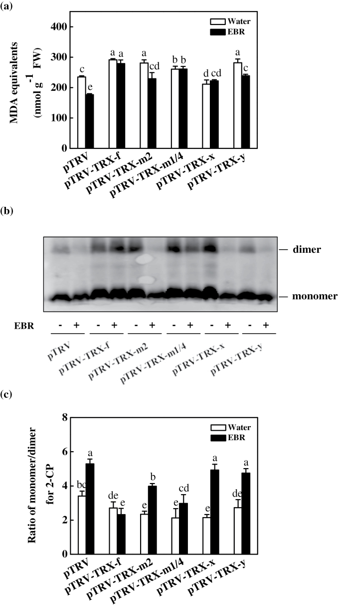
Fig. 4.
Changes in the lipid peroxidation and the redox state of 2-Cys peroxiredoxin (2-CP) protein in the leaves of virus-induced gene silencing (VIGS) plants as influenced by EBR application. (a) Changes in content of malondialdehyde (MDA) equivalents. (b) Changes in the redox state of 2-CP as investigated by non-reducing SDS–PAGE. The samples were separated by non-reducing SDS–PAGE and analysed in a western blot analysis with anti-2-CP. (c) The ratio of monomer/dimer for 2-CP from (b) as quantified by Quantity One. Leaf samples were taken at 24h after EBR treatment. Data are the means of four replicates with SDs. Means followed by the same letter are not significantly different according to Tukey’s test (P<0.05).

