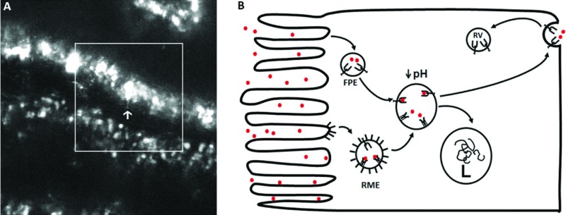
Fig. 2.
Transcytosis of albumin across proximal tubule cells in vivo using 2-photon microscopy. (A) Two-photon intra-vital time image taken in a Simonsen Munich Wistar rat given 2 mg of Alexa 568-RSA intravenously 24 hours before imaging shows vesicular and tubular structures containing albumin. A single frame with a large accumulation of albumin is indicated by the arrow; note the orientation of the apical membrane is opposite the arrow. The formation of a tubular structure extending from an intracellular compartment toward the basal pole of the PTC is shown at the end of the arrow. (B) Schematic of albumin entering into a PTC either via unbound in a fluid phase vesicle/endosome (FPE) or bound to megalin-cubulin as a receptor mediated endosome (RME) at the apical surface via a clatherin coated pit. With acidification to a pH of less than or equal to 6, the megalin-cubulin binding of albumin diminishes while that of FcRn increases dramatically. As such, there is an exchange of albumin from megalin-cubulin to binding to FcRn and this carrier then mediates transcytosis. When the transcytotic vesicle unites with the basolateral membrane, the increase in pH of the interstitial compartment releases albumin to diffuse into the interstitium and be transcytosed across endothelial cells again using FcRn as the carrier. FcRn then recycles to the apical membrane area via the recycling vesicle (RV). L stands for lysosome.

