
Figure 2.
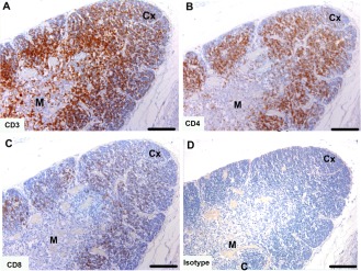
Immunohistochemical labeling with T cell subset markers in Tasmanian devil thymus. (A) anti‐CD3, (B) anti‐CD4 (C) anti‐CD8 and (D) isotype control. CD4+ T cells appear to be more prevalent than CD8+ T cells. Cx, cortex; M, medulla. Scale bar represents 100 μm.