
Figure 10.
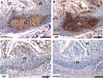
Immunohistochemical labeling with B cell subset markers in Tasmanian devil gut‐associated lymphoid tissue. (A) anti‐CD79b, (B) anti‐devil IgM, (C) anti‐devil IgG and (D) isotype control. Fl, follicle; Vi villus. Scale bar represents 100 μm.