
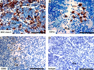
Figure 11.
Immunohistochemical labeling with dendritic cell markers in Tasmanian devil lymph node. (A) anti‐HLA‐DR (MHC class II), (B) anti‐CD1a (C) anti‐CD83 and (D) isotype control. Cx, cortex; Pcx, paracortex. Scale bar represents 100 μm.
Official websites use .gov
A
.gov website belongs to an official
government organization in the United States.
Secure .gov websites use HTTPS
A lock (
) or https:// means you've safely
connected to the .gov website. Share sensitive
information only on official, secure websites.
Immunohistochemical labeling with dendritic cell markers in Tasmanian devil lymph node. (A) anti‐HLA‐DR (MHC class II), (B) anti‐CD1a (C) anti‐CD83 and (D) isotype control. Cx, cortex; Pcx, paracortex. Scale bar represents 100 μm.