
Figure 14.
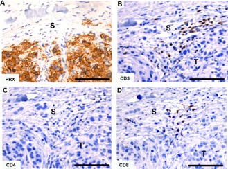
Immunohistochemical labeling with T cell markers in Tasmanian devil DFTD tumors. (A) anti‐periaxin (DFTD tumor cell marker), (B) anti‐CD3, (C) anti‐CD4 (no positive cells), (D) anti‐CD8. The isotype control is shown in figure 15. S, stroma; T, tumor. Scale bar represents 100 μm.