
Figure 4.
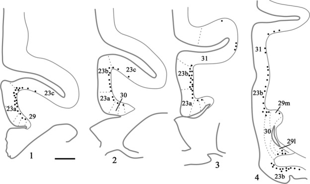
Drawings of four coronal sections depicting a case (CSR) with a single injection of DY placed in the medial dorsal nucleus. In this case each dot represents two labelled cells within the cortex. Section 4.1 is the most rostral. Scale bar, 1.5 mm.