
Figure 7.
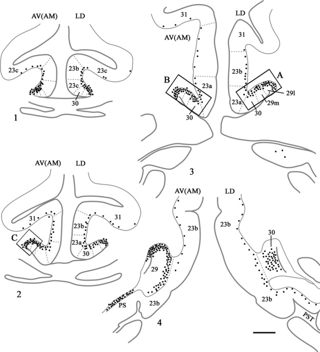
Distribution of retrograde label (Case BRh5) following an injection of FB into the caudal part of the anterior ventral nucleus in the left hemisphere, along with a second injection of FB into the laterodorsal thalamic nucleus in the right hemisphere. The dots reflect the relative distributions of labelled cells as there were too many to depict individually (see Fig.8). Photomicrographs of the three areas in boxes (A, B and C) are shown in Fig.8. PS, prosubiculum; PST, prostriate cortex. Scale bar, 2.0 mm.