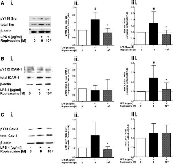
Figure 6.

Effect of ropivacaine on LPS-induced phosphorylation and expression of Src, ICAM-1, and caveolin-1 in cultured endothelial cells. Representative Western blots and quantitative densitometry analysis of human lung microvascular endothelial cell (HLMVEC) lysates show (A) Src, (B) ICAM-1, and (C) Cav-1 after treatment with LPS (4 μg/ml) in absence or presence of ropivacaine at a concentration of 1 nM (10−9 M) for 4 hours. Quantitative densitometry analysis show (Aii, Bii, Cii) the ratio of phosphorylated over total protein or (Aiii, Biii, Ciii) total protein over β-actin in cells treated with LPS (4 μg/ml) in absence (black bar) or presence (striped grey bar) of ropivacaine compared to untreated cells (white bars, set as 1.0). All values shown are mean ± SD (n = 6-11/group). #p < 0.05 vs. untreated cells (control), *p < 0.05 compared to LPS alone.
