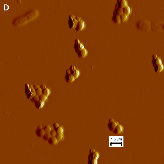
Figure 7.

Atomic force microscopy of P. aeruginosa and MRSA in mono- and co-culture on gelatin-coated mica. (A & B) Showing overview of P. aeruginosa BK-76 mono-culture and two individual cells with flagella (C) showing MRSA M05-35 cell aggregation in mono-culture (D & E) showing co-culturing of P. aeruginosa BK-76 + MRSA M05-35. (D) Shows an overview of the co-culture with P. aeruginosa BK-76 single and attached cells. (E) Shows two attached P. aerugionsa BK-76 cells ‘attacking’ a single MRSA M05-35 cell with the appearance of spilled cytoplasmic contents.
