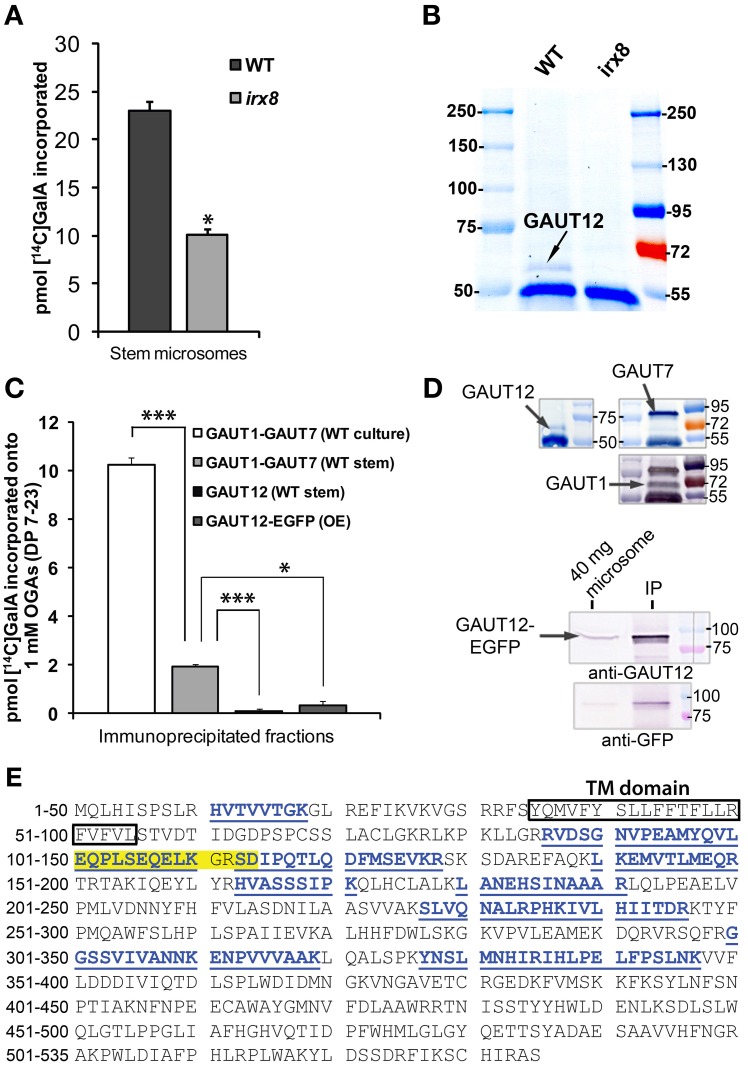
Figure 9.
Enzymatic activity assays for GAUT12 and the anti-GAUT12 antibody. (A) HG:GalAT activity of total microsomes (50 μg) from WT and irx8-5 stems. Value = mean ± standard deviation (n = 3), *indicates significant reduction of HG:GalAT activity in irx8 stem microsomes at P(t-test) < 0.05. (B) Western blot analysis of immunoprecipitated (IP) GAUT12 fractions from wild-type (WT) and irx8-5 7-week-old stem microsomes (500 μg total protein) using 30 μl anti-GAUT12 antibody-conjugated magnetic beads. GAUT12 protein band as indicated (~58–60 kDa) is present in WT but absent in irx8-5 microsomes. (C) HG:GalAT activity of immunoabsorbed-GAUT12 from WT stem microsomes and from GAUT12-EGFP fusion protein expressed in Arabidopsis suspension culture cells (OE). Immunoabsorbed-GAUT1:GAUT7 complex from WT suspension cell cultures and from WT stem microsomes were used as positive controls. Equal amounts of the corresponding antibody-conjugated beads (30 μl) were incubated with equal amounts of TX-100 permeabilized microsomes from different tissues (500 μg total protein). Value = mean ± standard deviation (n = 3), *, *** indicates significant reduction of HG:GalAT activity determined by One-Way ANOVA and post-hoc Bonferroni corrected t-test at p < 0.05 and p < 0.001, respectively. Immunoabsorbed-GAUT12 and GAUT12-EGFP activity are similar to background with P(t-test) values = 0.44 and 0.22, respectively. All enzyme activity assays were repeated at least twice. One set of representative results is shown. (D) Western blots showing the presence of GAUT12, GAUT1, and GAUT7 in the corresponding immunoabsorbed fractions from WT stem tissues. GAUT12 was immunoabsorbed by the anti-GAUT12 antibody, and the GAUT1:GAUT7 complex was immunoabsorbed by the anti-GAUT7 antibody. Arabidopsis culture expressed-GAUT12-EGFP (~86 KDa) fusion protein in the anti-GAUT12 immunoabsorbed fractions used in reactions in (C) shown in the lower panel. (E) GAUT12 protein sequence (535-a.a.). The antigenic-peptide used to generate the anti-GAUT12 antibody is highlighted in yellow. Peptides identified in GAUT12-IP fractions by LC-MS/MS are labeled in blue with underlines. Boxed sequence indicates the GAUT12 transmembrane (TM) domain predicted by TmHMM_v2.

