
Figure 3.
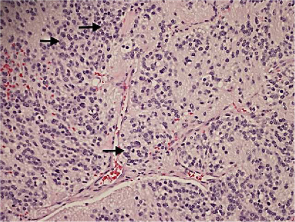
Resected mediastinal mass surgical pathology. Hemotoxylin & Eosin stained section of resected mediastinal mass reveals scattered small Homer-Wright rosettes (arrows): small dark neuroblastoma cells in circular groups around pale fibrillary neuropil (200× magnification).