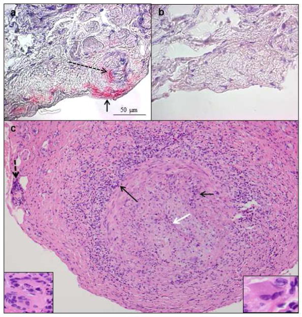
Fig. 4.
VZV antigen in the temporal artery of a patient with giant cell arteritis. Immunohistochemistry revealed VZV antigen (a red color) in the adventitia (arrow) and its nerve bundles (dashed arrow), as well as in media and intima (not shown) after staining with anti-VZV antibody; no staining was seen using anti-HSV-1 antibody (b) or normal rabbit serum (not shown). 600X. Hematoxylin & eosin staining of cross-section of the temporal (c) revealed extensive inflammation in the adventitia (long black arrow), as well as in the media (short black arrow) and vaso vasorum (dashed black arrow). White arrow spans the thickened intima and points to an occluded lumen. Numerous multinucleated giant cells were seen throughout the artery (insets). 200X. (Permission granted from Journal of Neurological Sciences)

