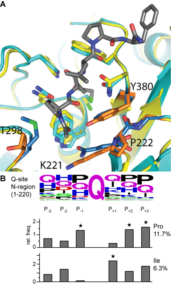
Figure 10.

Model of the AgTG3 active site. (A) Homology model of AgTG3 (yellow) superimposed with TG2-inhibitor complex 2Q3Z (cyan). Non-conserved AgTG3 residues (K221, P222, T298, Y380) are shown (orange) in comparison to TG2 (blue) in the peptide-binding groove. (B) Proline is highly enriched at the P-1 relative to Gln in the N-terminal region of Plugin, justifying the bias towards cyclic amino acid variants in the initial library.
