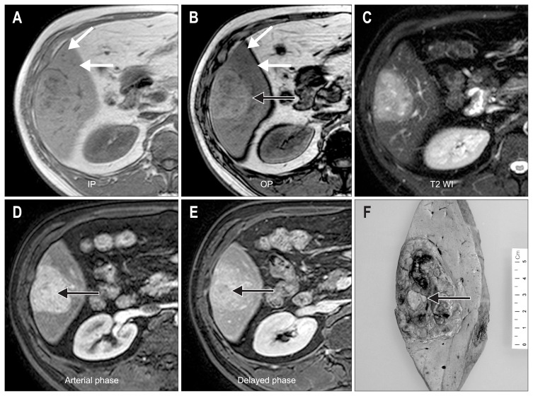
Fig. 1.
(A–E) The radiologic features of case 1. In-phase (IP) (A) and opposed-phase (OP) (B) T1-weighted, gradient echo images show an iso-intense lesion on IP images, whereas a hyperintense mass (black arrow) appears on the OP image due to decreased signal in the surrounding liver (white arrows). A T2-weighted image (T2 WI) (C) shows a slightly hyperintense mass. An axial, gadobenate dimeglumine-enhanced, T1-weighted, gradient echo image in the arterial phase (D) shows a homogeneous enhancement of the mass (arrow). An axial, gradient echo image in the delayed phase (E) shows no washout of the contrast. These features are consistent with hepatocellular adenoma; however, a washout area within the enhanced mass is suspicious for hepatocellular carcinoma (arrow). (F) Gross features of case 1. The resected specimen demonstrates a well-defined and multilobular mass measuring 7×5 cm, and a yellowish nodule is noted at the center (arrow).

