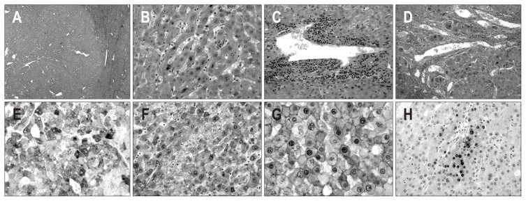
Fig. 2.
Microscopic and immunohistochemical features of case 1. (A) The mass shows a well-defined border with an expanding growth pattern (H&E stain, ×40). (B) The tumor cells of the outer nodule show a trabecular pattern of one- or two-cell thickness with mild nuclear atypia (H&E stain, ×200). (C) Several abnormally shaped blood vessels with infiltration of mononuclear inflammatory cells are noted within the tumor (H&E stain, ×200). (D) The tumor cells of the inner nodule show stromal invasion (H&E stain, ×200). (E, F) The tumor cells of the outer and inner nodules show diffuse and strong cytoplasmic expression of serum amyloid protein A (E) and glutamine synthetase (F) (×200). (G) The tumor cells of the outer and inner nodules show diffuse nuclear expression of β-catenin (×400). (H) The tumor cells of the inner nodule show the focal expression of heat shock protein 70 (×200).

