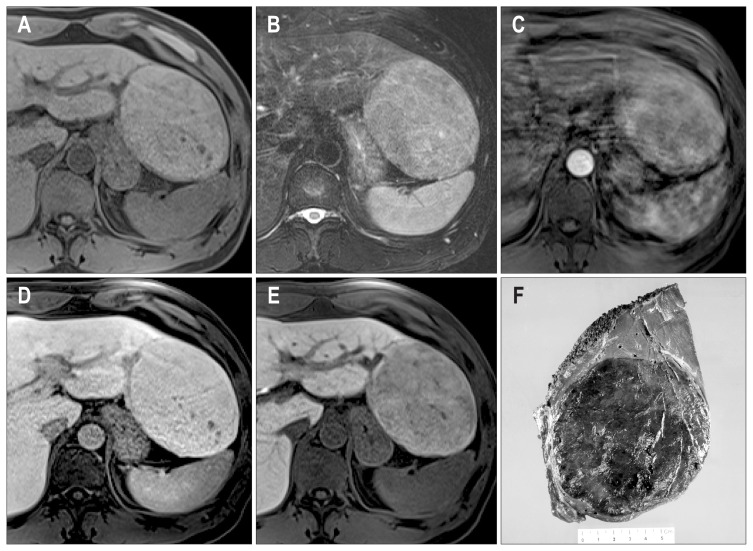
Fig. 5.
(A–E) Gadoxetic acid enhanced magnetic resonance imaging of case 3. There is a 10-cm mass lesion in the left lateral segment with an isointense signal in the T1-weighted image (A), diffuse high signal intensity and several bright foci in the T2-weighted image (B), arterial enhancement with slight washout (D), and poor contrast uptake on the hepatobiliary phase image (E). (F) The gross features of case 3. The cut surface of the mass is well-delimited and dark green in color with focal areas of congestion and hemorrhage.

