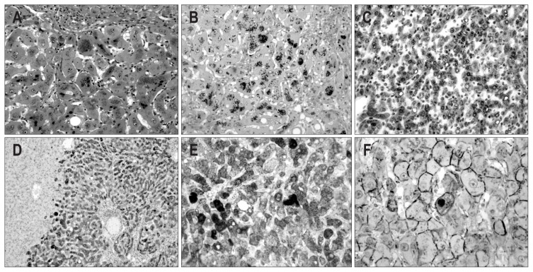
Fig. 6.
Microscopic and immunohistochemical features of case 3. (A) The tumor cells are composed of hepatocyte-like cells with mild atypia. Dark brown granular pigment is present in the cytoplasm of the tumor cells (H&E stain, ×200). (B) The pigment is positive by Fontana-Masson staining (×200). The cytoplasm of the tumor cells is diffusely positive for serum amyloid protein A (C, ×200), C-reactive protein (D, ×100), and glutamine synthetase (E, ×200). The tumor cells show the focal nuclear expression of β-catenin (F, ×400).

