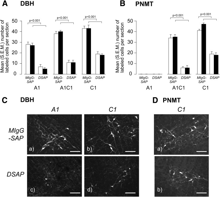
Figure 2.
Animals injected in the PVH with a DSAP show a significant loss of DBH (A and C) and PNMT (B and D) immunoreactive neurons in the A1, A1/C1, and C1 regions of the VLM compared with animals injected with a MIgG-SAP. Animals were exposed to either rapid- (open bars) or slow-onset (solid bars) hyperinsulinemic-hypoglycemic clamp. Results in A and B are expressed as mean (SEM) number of labeled cells per section. C and D show DBH and PNMT in the A1 (DBH only) and C1 regions of the VLM. Scale bar = 100 μm.

