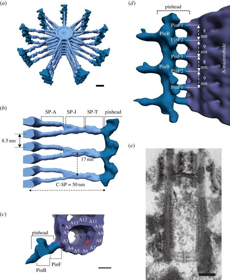
Figure 3.
Cryo-electron tomography images of the cartwheel in Trichonympha. (a) Three-dimensional representation of a stack of cartwheels (light blue) with pinheads (dark blue). Each cartwheel is composed of nine approximately 50 nm long spokes emanating from the central hub (diameter: approx. 22 nm). Scale bar, 10 nm. (b) Side view of the cartwheel stack highlighting the cartwheel spoke (C-SP). Hubs (left-hand side) are stacked with an 8.5 nm periodicity. Spokes in the adjacent cartwheels are paired and merged at approximately 20 nm from the hub. The spoke is structurally divided into a spoke arm (SP-A), a spoke junction (SP-J) and a spoke tip (SP-T). (c) Top view of the pinhead and the A-tubule of the triplet microtubule (violet). The pinhead is subdivided into a pinbody (PinB) and pinfeet (PinF) that connect the pinbody to the microtubule. Scale bar, 10 nm. (d) Side view of the pinhead associated with the microtubule. The pinfeet consist of pinfoot 1 (PinF1) and pinfoot 2 (PinF2), which alternate every 8 and 9 nm along the microtubule axis. The tilting of the pinfeet towards the proximal end of the centriole defines the polarity of the cartwheel. Reproduced with permission from [21]. Copyright 2013 Elsevier. (e) A thin section image of the Chlamydomonas centriole. The cartwheel stack displays approximately 20 nm periodicity, a distance close to that of merged spokes observed in the Trichonympha cartwheel. Scale bar, 100 nm. (Online version in colour.)

