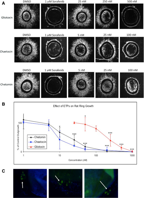
Figure 1.
Rat ring assay shows dose-dependent inhibition of microvessel outgrowth by ETPs. A, Cells were treated with DMSO (negative control), 1 μM sorafenib (positive control), and increasing concentrations of gliotoxin, chaetocin, and chetomin. Three concentrations of each ETP are shown to illustrate the dose-dependent effect of the drugs on microvessel outgrowth. B, Dose response curves showing a decrease in outgrowth with increasing ETP concentrations. Gliotoxin, chaetocin, and chetomin had a GI50 of 151, 8, and 20 nM, respectively. Data points are presented as mean S.E.M (error bars) from independent experiments run in triplicate (n = 6). *, p < 0.05, **, p < 0.001, ***, p < 0.0001. C, Rat aortic rings at day 5 were stained with a HIF-1α monoclonal primary antibody and a FITC-conjugated secondary antibody to label endothelial cells. The merged image shows DAPI-stained nuclei in blue and HIF-1α expression in green (indicated by arrows). Three representative images are shown.

