Figure 3.
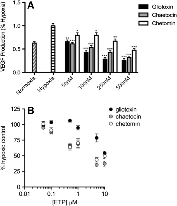
ETPs decrease VEGF secretion in a dose-dependent manner. A, Hypoxia was induced for 18 h in PC3 cells in the absence or presence of the indicated concentrations (1000 nM is not shown) of gliotoxin, chaetocin, and chetomin. This was followed by ELISA quantification of secreted VEGF normalized to DMSO under hypoxic conditions. A repeated measures ANOVA was performed on the data; Hochberg’s post-hoc method was used to adjust the p-values. Data are presented as mean ± S.E.M from independent experiments run in triplicate (n = 2-7). *, p < 0.05, **, p < 0.001, ***, p < 0.0001. B, PC3 cells were treated with increasing concentrations of ETPs or with vehicle control (DMSO). Plates were placed in either a normoxic incubator or hypoxic chamber for 18 h. Cell viability was then determined using the CellTiter-Blue cell viability reagent. Data points are presented as mean ± S.E.M from independent experiments run in triplicate (n = 4).

