Figure 1.
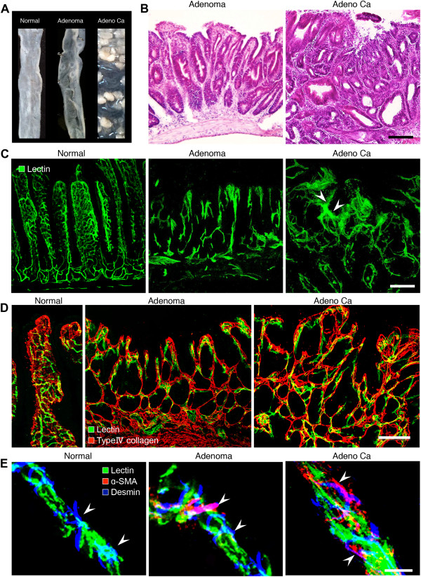
Relationship between tumor progression and local microvascular changes during multistep carcinogenesis in ApcMin/+ mice. A, Representative images of normal intestine or intestinal polyps: normal region (left), adenoma (center), adenocarcinoma (right). n = 20. B, H&E staining of adenoma and adenocarcinoma. Scale bars: 100 μm. C, Tomato lectin-labeled vascular architecture in a normal region, adenoma, and adenocarcinoma. Note the marked structural abnormalities in the tumor vessels, including altered vascular density, vessel compression, uneven diameter, blind ending vessels, and leakiness (arrowheads) in adenocarcinoma. Blood vessels in adenoma and adenocarcinoma lost their vascular hierarchy. Scale bars: 100 μm. D, Double-staining for FITC-tomato lectin and type IV collagen in a normal region, adenoma and adenocarcinoma during the adenoma-carcinoma sequence. Scale bars: 100 μm. E, Pericyte distribution in tumor vessels during the adenoma-carcinoma sequence in ApcMin/+ mice. Vessels in a normal region of the small intestine, adenomas, and adenocarcinomas. Vessels were stained with FITC-tomato lectin (green). Pericytes were visualized with a combination of α-SMA (red) and desmin (blue). Scale bars: 20 μm.

