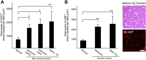
Figure 3.

Comparison of vascular density and angiogenic patterns between mouse intestinal polyps and human surgical specimens. A, Vascular density and angiogenic patterns of normal intestine and tumor tissues in ApcMin/+ mice. The difference in density of microvessels per field between normal tissue and adenoma or adenocarcinoma in ApcMin/+ mice was compared by measuring the total area comprising vessels at each stage. The MVD was estimated by measuring the total areas of normal blood vessels and newly formed blood vessels in 10 separate fields of normal small intestine, mouse adenoma, or mouse adenocarcinoma at a magnification of 600 ×. *P < 0.05, **P < 0.01. B, Vascular density and angiogenic patterns of normal human intestine and colon tumor specimens. The MVD was estimated by measuring total areas comprising blood vessels in 10 separate fields of normal colon, human adenoma, or human adenocarcinoma at a magnification of 600 ×. Hematoxylin and eosin (H&E) staining of adenocarcinoma in human specimens and staining for vWF, a marker for human endothelial cells, in adenocarcinoma. **P < 0.01. Scale bar: 100 μm.
