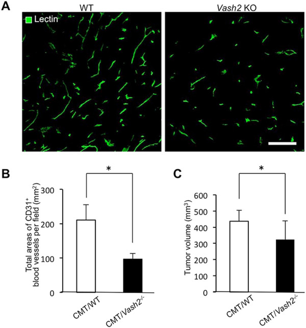
Figure 5.
Tumor progression and tumor angiogenesis in Vash2 KO mice. A, Tomato lectin staining of tumor vessels in CMT93 tumor-bearing WT mice (left) and Vash2-/- mice (right). Scale bars: 100 μm. Compared with CMT93 tumors in WT mice, the number of tumor vessels markedly decreased in tumors of Vash2-/- mice. B, Microvascular density (MVD) was estimated by measuring the total area comprising CD31+ blood vessels in three separate fields of CMT93 tumors in WT mice or Vash2-/- mice at 400× magnification. *P < 0.05, n = 8. C, Growth of CMT93 tumors transplanted into WT or Vash2-/- mice. Open columns represent CMT93 tumor-bearing WT mice and closed columns represent CMT93 tumor-bearing Vash2-/- mice. *P < 0.05, n = 15.

