
Figure 2.
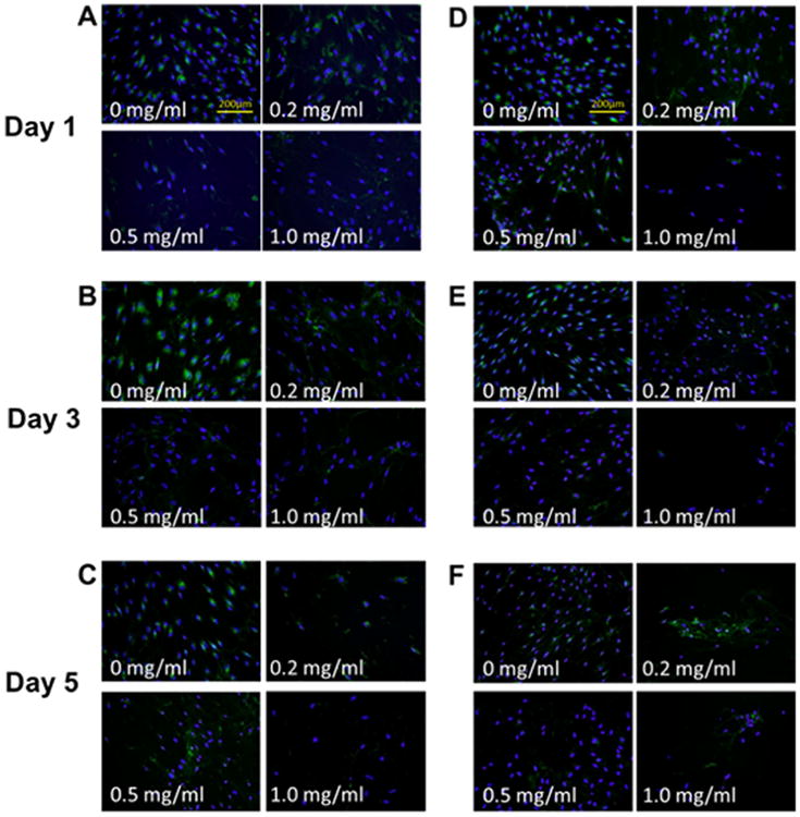
Collagen type I expression of fibroblasts after mitomycin C treatments. (A–C) Representative immunocytochemistry (ICC) staining of normal vocal fold fibroblasts (nVFFs) over 5 days after various doses of mitomycin C administration (original magnification ×200). (D–F) Representative ICC staining of scarred vocal fold fibroblasts (sVFFs). Collagen type I was stained green and cell nuclei was stained blue. [Color figure can be viewed in the online issue, which is available at wileyonlinelibrary.com.]