
Figure 4.
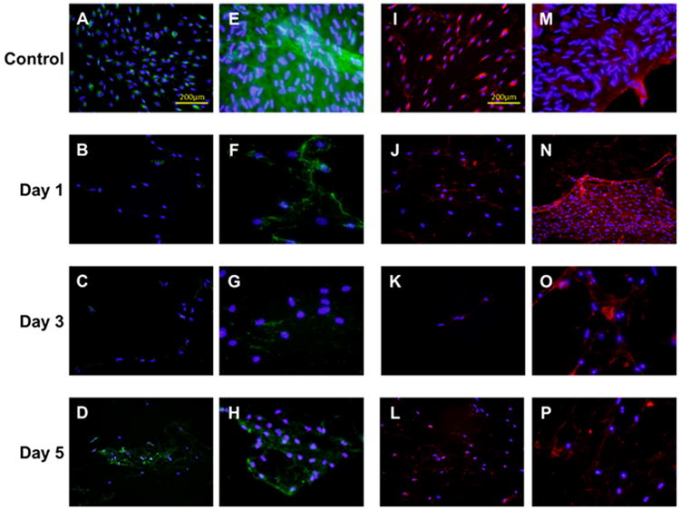
Collagen type I and III expression of scarred vocal fold fibroblasts (sVFFs) after mitomycin C treatments in the validation experiment (original magnification ×400). All cells were treated with 1.0 mg/ml mitomycin C except the serum controls. (A–H) Representative immunocytochemistry (ICC) staining of collagen I (A–D) with cell membrane permeabilization (E–H) in the absence of permeabilization. (I–P) Representative ICC staining of collagen III (I–L) with cell membrane permeabilization (M–P) in the absence of permeabilization. Collagen type I and III was stained green and red respectively. Cell nuclei were stained blue. [Color figure can be viewed in the online issue, which is available at wileyonlinelibrary.com.]