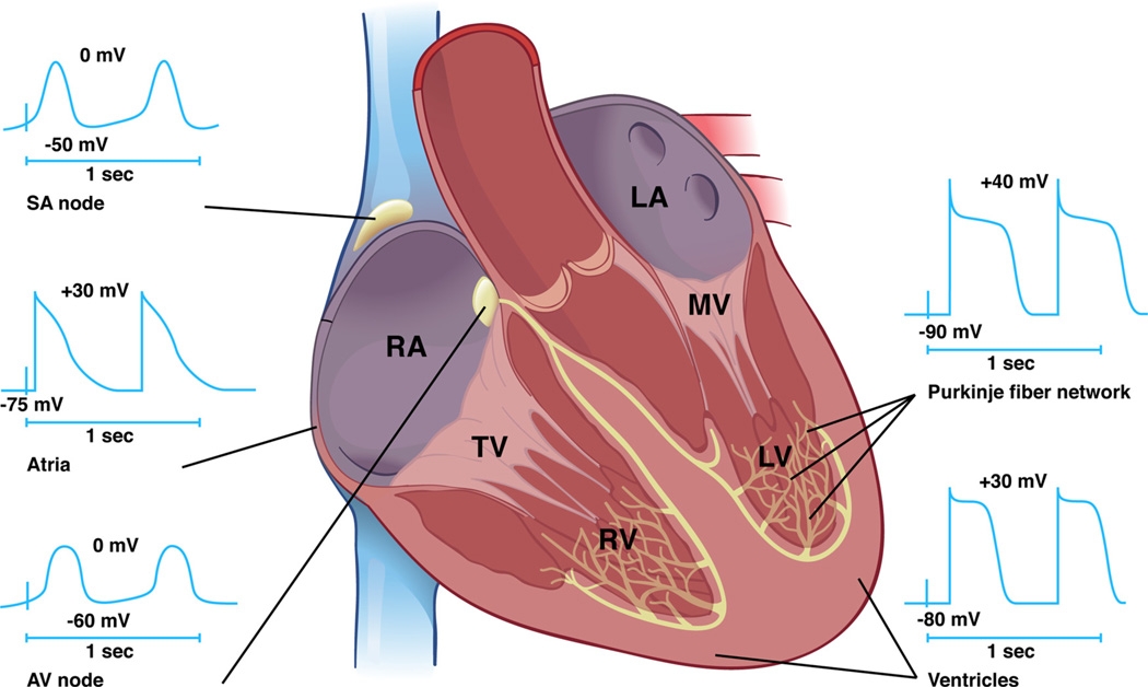
Figure 1. Functional anatomy of the cardiac conduction system.
The cardiac impulse is initiated in the SAN and propagates through the atria. The electric impulse is then delayed in the AVN before rapid conduction through the right and left bundle branches before terminating in the Purkinje fiber network, which directly interfaces with ventricular cardiomyocytes. Distinct time-voltage relationships are noted in the SAN, atria, AVN, PFs, and ventricles. RA indicates right atrium; LA, left atrium; TV, tricuspid valve; MV, mitral valve; RV, right ventricle; LV, left ventricle.

