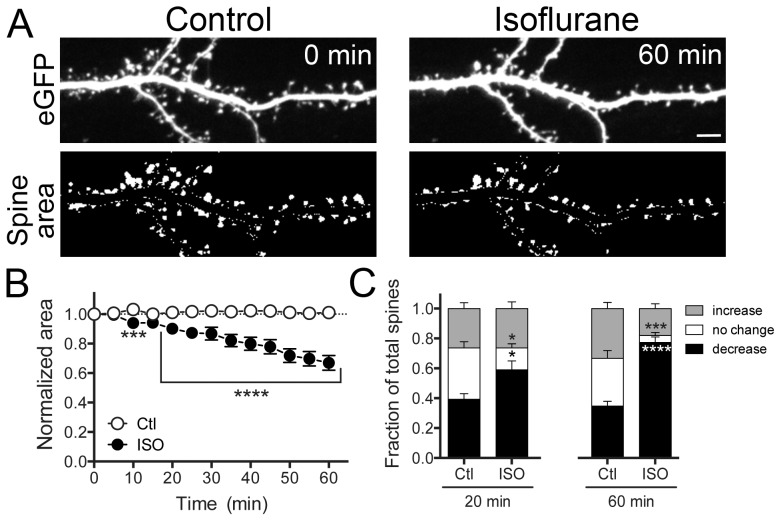
Figure 2. Isoflurane reduces dendritic spine area.
Hippocampal neuron cultures transfected with eGFP were exposed to 95% air/5% CO2 (Ctl) or 2 vol% isoflurane in 95% air/5% CO2 for 60 min at 37°C. Representative images show eGFP fluorescence (A-top panels) and thresholded overall spine area after manual dendrite subtraction (A-bottom panels). A time-dependent decrease in total spine area was observed (B) by two-way ANOVA with Sidak post hoc test (***p<0.001). Changes in individual spine area of >10% (increase) or <10% (decrease), or no change in area were evident at 20 and 60 min (C) by two-way ANOVA with Sidak post hoc test (*p<0.05; ***p<0.001; ****p<0.0001). Data are mean ± SEM; n = 1500 to 1800 dendritic spines per experimental group. Scale bar = 5 µm.

