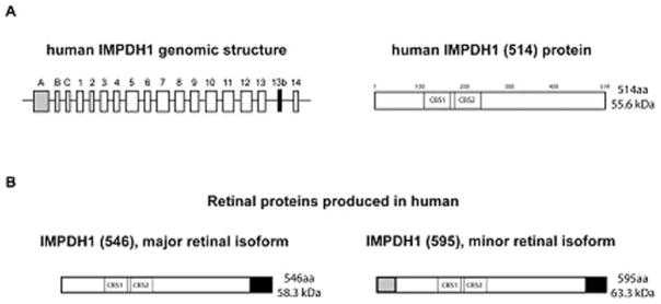
Fig. 62.2.

(a) Genomic structure of IMPDH1 in humans and the ‘canonical’ IMPDH1 (514) protein. The ‘canonical’ IMPDH1 (514) protein results from translation of exons 1–14. (b) Retinal-specific isoforms of IMPDH1 in human. The black portion of the proteins represents the amino acid sequence resulting from the addition of exon 13b in the spliced transcript. The grey portion of the protein represents the sequence resulting from the addition of exon A in the spliced transcript. IMPDH1 (546) is the most abundant isoform found in human retina and results from the translation of exons 1–14 and exon 13b. The less abundant isoform in human retina is IMPDH1 (595) which is translated from exons 1–14 and exons A and 13b. CBS1 and CBS2 illustrate the approximate location of the cystathionine β-synthase-like (CBS) domains in the proteins
