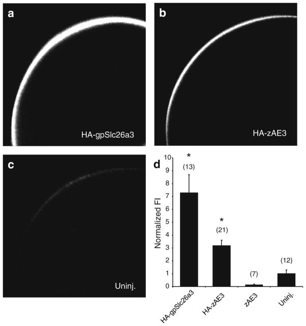
Fig. 4.
HA-zAe3 is expressed at or near the oocyte surface. Xenopus oocytes (diameter 1–1.3 mm) expressing HA-tagged zebrafish Ae3 show significantly more protein surface expression than uninjected oocytes. Confocal immunofluorescence images of representative median intensity showing uninjected oocytes (Uninj.) or oocytes previously injected with cRNA encoding HA-tagged zAe3 (50 ng, HA-zAE3), untagged zAe3 (50 ng, zAE3) or HA-tagged guinea pig Slc26a3 (50 ng, HA-gpSlc26a3). The bottom right panel shows mean normalized fluorescence intensities for (n) oocytes. * p<0.05 vs. zAe3 and uninjected

