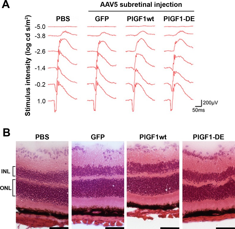
Figure 3. .
ERG and histochemical analyses of retinas after AAV5 delivery. Intravitreous administration (4 animals per group) of AAV5-GFP, AAV5-PlGF1wt, or AAV5-PlGF1-DE did not induce retinal damage as monitored by ERG and histochemical analysis, compared to PBS. (A) Representative wave form and amplitude responses during scopic flash ERG. (B) Representative pictures of H&E-stained sections illustrating the normal retina morphology. ONL thickness (expressed in μm ± SEM), PBS 52.5 ± 2.3, AAV5-GFP 53.4 ± 1.6, AAV5-PlGF1wt 52.8 ± 2.6, AAV5-PlGF1-DE 49.4 ± 4.8. INL thickness (expressed in μm ± SEM), PBS 31.3 ± 4.1, AAV5-GFP 30.6 ± 3.5, AAV5-PlGF1wt 29.3 ± 3.7, AAV5-PlGF1-DE 30.2 ± 3.2. Scale bar: 50 μm.

