
Figure 5.
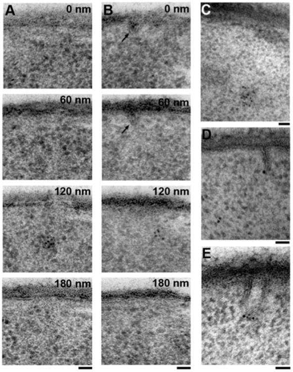
Identification of eisosomes by anti-GFP labeling of a strain expressing GFP-tagged Pil1p, the most abundant eisosomal/MCC component. A: A 60 nm section series showing the most commonly observed pattern of Pil1p-GFP labeling under our conditions of exponential growth, where dense clusters of label are neither at the plasma membrane nor associated with any visible deformation of the plasma membrane. B: A 60 nm section series showing a cluster of Pil1p-GFP label in proximity to a shallow furrow (arrow). C: Cluster of Pil1p-GFP label 300 nm away from the plasma membrane. D: Pil1p-GFP label associated with the tip of a rare, 100 nm deep, sheet-like furrow, as previously described (Stradalova et al., 2009). E: Pil1p-GFP label at the tip of another obliquely sectioned, sheet-like furrow. Scale bars = 50 nm.