
Figure 3. EMR1 expression on tissue eosinophils.
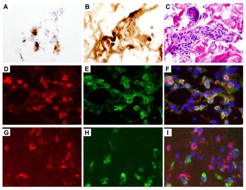
A-C, Skin biopsy from a subject with HES. Frozen section stained with antibody to EMR1 (A). Adjacent paraffin-embedded sections stained with antibody to EPX (B) and with H&E (C) showing eosinophilic infiltration and degranulation. The arrows indicate representative eosinophils (C). (B). Original magnification 600x (A, B,) and 100x (C). D-I, Nasal polyp biopsy. Immunofluorescence staining of EMR1 in red (D,G), EPX (E) or CD68 (H) in green, and overlay (F,I) showing EMR1 staining on eosinophils and not on macrophages. Original magnification 640x.