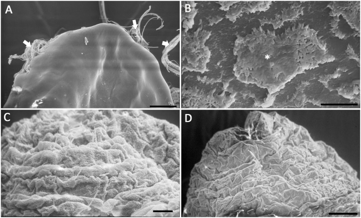
Figure 4. Scanning electron micrographs of the surface morphology of M. matsumurae at different developmental stages.
A, 2nd-instar nymph, showing the insect body was smooth with wax filaments secreted from spiracles on both sides. Scale bar = 100 µm; B, magnified view of the 2nd-instar nymph, showing the thin wax layer on the insect surface. Scale bar = 10 µm; C and D, male 3rd-instar nymph and adult female, showing both of their body segments are distinct with obvious intersegmental folds. C, Scale bar = 20 µm. D, Scale bar = 100 µm.

