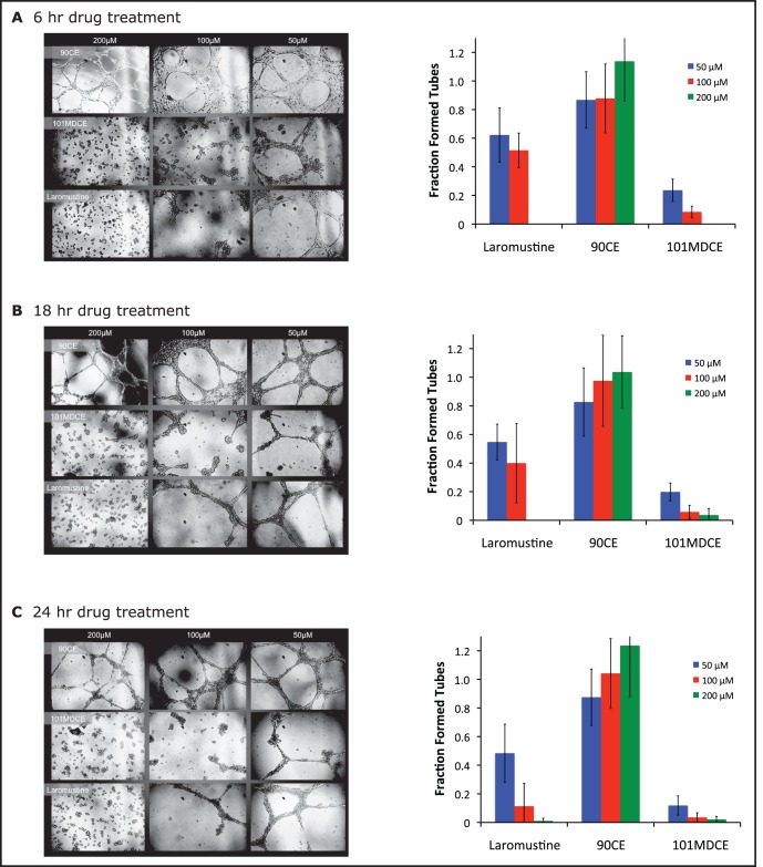
Figure 6. Inhibition of EC tube formation in vitro by 101MDCE.
Human umbilical vein endothelial cells (HUVECs) were treated with 50 µM, 100 µM, or 200 µM Laromustine, 90CE, or 101MDCE (or DMSO control). 3×10⁴ cells/cm2 were plated onto 96-well plates containing a thin film of Matrigel (BD Bioscience). The cells were examined at 6 hrs (A), 18 hrs (B), and 24 hrs (C) using a phase contrast inverted microscope. Tube structures were quantified by placing the microscope over the center of the well and counting fully formed tubes and partially formed tubes (defined as more than three-quarters closed) within view. Tube formation was normalized in each case to that of the DMSO control.

