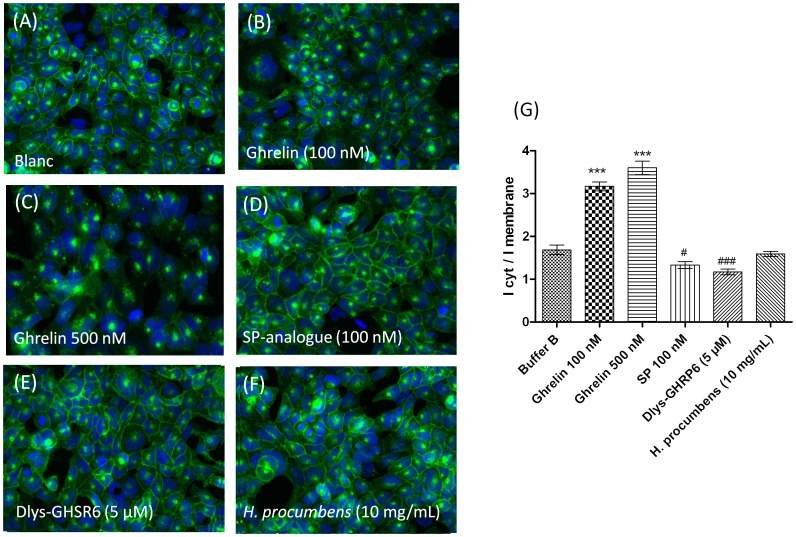
Figure 4. H. procumbens root extract does not internalize the GHS-R1a receptor.
Hek cells stably expressing the GHS-R1a receptor as a C-terminal EGFP fusion protein were visualized using the IN Cell Analyser 1000 (GE Healthcare) following different treatments: untreated (A), ghrelin (B,C), [D-Arg1, D-Phe5, D-Trp7,9, Leu11]-substance P (SP-analogue) (D), (Dlys3)-GHRP-6 (E) and H. procumbens root extract (F) at the indicated concentrations for 1 h at 37°C. Ligand-mediated GHS-R1a-EGFP translocation is quantified following the EGFP fluorescent trafficking away from membrane into vesicles within the cytosol. Graph represents the mean ± SEM of the fluorescence intensity of perinuclear receptor expression versus plasma membrane receptor expression from a representative experiment out of two independent experiments with each treatment performed in triplicate (G). Significant increased internalization is depicted as *** p<0.001, and significant decreased internalization is depicted as ##p<0.01, #p<0.05 with respect to internalization obtained from assay buffer (blanc).

