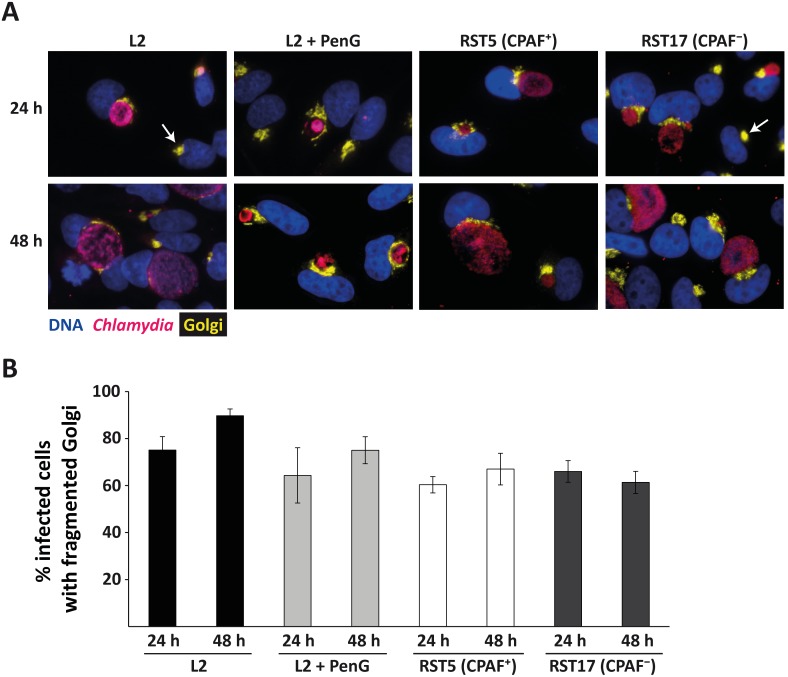
Figure 2. Golgi-fragmentation during acute and persistent infection.
(A) YFP-Golgi-HeLa cells were infected with C. trachomatis L2 with or without addition of 100 U/ml PenG, RST17 (CPAF−) and the corresponding control strain RST5 (CPAF+) [14], incubated for the indicated times and processed for immunofluorescence. Blue: Hoechst DNA-stain, yellow: GA, pink: Chlamydia. All images were taken at 40-fold magnification. Arrows point to uninfected cells displaying a normal, not fragmented GA. (B) Quantification of the portion of infected cells showing a fragmented GA. All infected cells as well as all infected cells with fragmented GA were counted and the ratio of fragmentation-positive cells was calculated (number of infected cells counted: 499, 629, 478, 517, 397, 475, 380, 522 from left to right). Shown are means of 3 independent experiments ± SEM.

