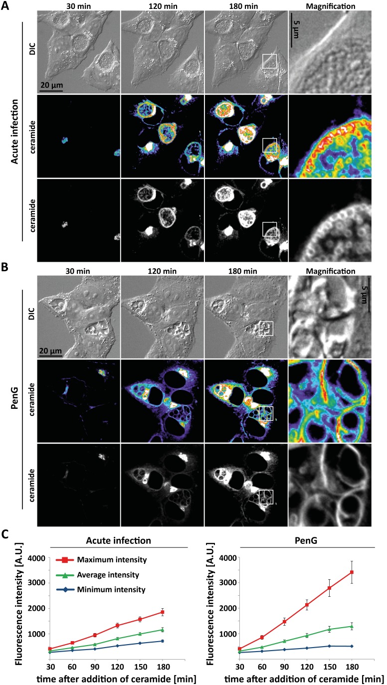
Figure 3. Ceramide transport in acute and persistent infection.
Hela cells were infected with C. trachomatis L2 at an MOI of 1 for 24 h without (A) or with addition of 100 U/ml PenG (B). Confocal microscopic movies were recorded and exemplary pictures taken 30, 120 and 180 minutes after addition of BODIPY-FL C5 ceramide are shown. The right column gives enlargements of the area indicated by the square. The heatmap ranges from purple (low intensity) to white (high intensity). (C) Quantification of fluorescence intensity. Averages of two independent experiments are given ± SEM (35 inclusions in acute and 26 inclusions in persistent infection).

