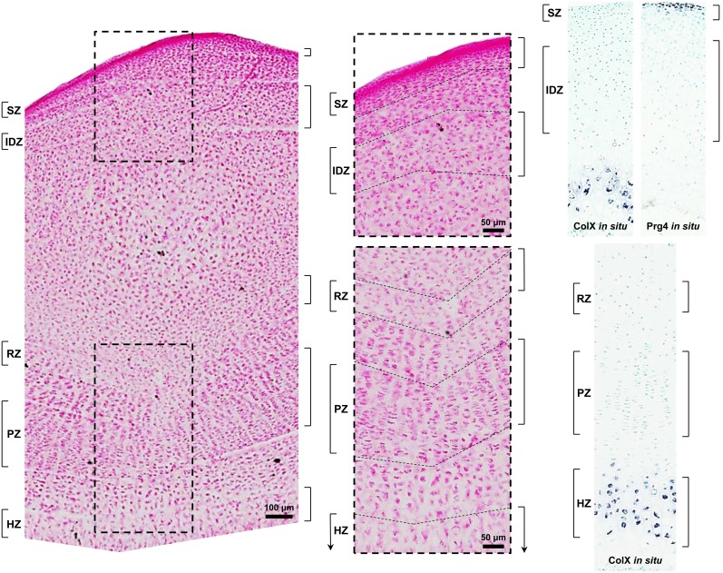
Figure 1. Photomicrograph of a longitudinal section of 10-day-old rat proximal tibia stained with eosin for manual microdissection.
Superficial (SZ) and intermediate/deep (IDZ) zones of articular cartilage and resting (RZ), proliferative (PZ), and hypertrophic (HZ) zones of growth plate cartilage were isolated with a razor blade. To minimize cross-contamination, a segment of cartilage between zones was discarded. Higher magnification is shown in the middle panel with respective regions delineated by dashed lines. For microarray analysis, only SZ, IDZ, and RZ were used, whereas all zones were used for real-time PCR. In situ hybridization of the articular cartilage SZ marker, Prg4, and the hypertrophic chondrocyte marker, Col10a1 (ColX), was used as a visual guide for manual microdissection. Hybridization was detected using NBT/BCIP substrates (purple) and tissues were counterstained with methyl green.

